Magnetic Resonance Imaging (MRI) at University Radiology Group

MRI vs. Other Imaging Tests | Specialty MRI Types | Preparation Instructions | What To Expect During and After

Magnetic Resonance Imaging (MRI) is a noninvasive diagnostic test that uses magnetic fields and radio waves to visualize structures within the body. MRIs can be used to see organs, muscle tissue, blood vessels, and tendons with improved clarity and resolution.

MRI vs. Other Imaging Tests

  • MRI vs. X-ray: Unlike X-rays, MRI scans do not use ionizing radiation and focus on soft tissues, rather than bones.
  • MRI vs. CT: Both MRIs and Computed Tomography (CT) scans can be performed with or without contrast material. MRIs often provide a clearer picture than a CT scan, but take significantly longer to perform. CT scans are preferred for acute traumas where speed is important.
  • MRI vs. Ultrasound: Ultrasounds offer a real-time, highly accessible way to assess soft tissue issues. When an ultrasound shows evidence of a mass, injury, or other condition, an MRI is often ordered to provide more detailed information.

Specialty MRI Types

MR Angiography (MRA)

MRA is an MR scan that looks at the blood vessels. This scan is usually done with the injection of a contrast agent. It is usually performed to find aneurysms, atherosclerosis, and/or abnormal narrowing in blood vessels.

MR Cholangiography (MRCP)

An MRCP is an MRI study of the gallbladder, biliary system and pancreas. It helps doctors evaluate patients for gallstones, pancreatic abnormalities and bile duct abnormalities.

MR Arthrography

MR Arthrography involves the injection of contrast into a joint followed by an MR scan of the joint. It may be performed using X-ray or ultrasound guidance. MR Arthrography can be used in place of non-contrast MRI, and is recommended for certain types of injuries or if the patient has had prior knee surgery.

MultiParametric MRI of the Prostate (mpMR)

This specialized MRI is used to identify prostate cancer, without the need for blind biopsy. It also helps physicians with cancer staging, which is important to developing a care plan.

MR Spectroscopy

MR Spectroscopy is a special type of MR imaging that uses additional scans to help determine the specific tissue types that comprise a mass or lesion. It is usually used for mass characterization in the central nervous system, including the brain and spinal cord.

MRI Preparation Instructions

Before your MRI, you’ll need to complete paperwork and answer questions about any metal present in your body. This is because the machine uses powerful magnets, which can cause injury by attracting certain metallic items. Make sure to note if you have any of these:

  • Cochlear implant or hearing aid
  • Pacemaker
  • Defibrillator
  • Neurostimulators
  • Permanent jewelry
  • Some medical stents and clips
  • Foreign bodies (such as shrapnel fragments)

Most joint replacements and metal supports used in orthopedic cases are made of MRI-safe materials, but you should still tell our team about any metal rods, nails, or joints you may have. You should also let our office know if you have any renal issues, or have ever had an allergic reaction to a contrast dye.

What To Expect During Your MRI

When you come to our office for your MRI, you’ll need to remove any clothing with metal in it, as well as all jewelry.

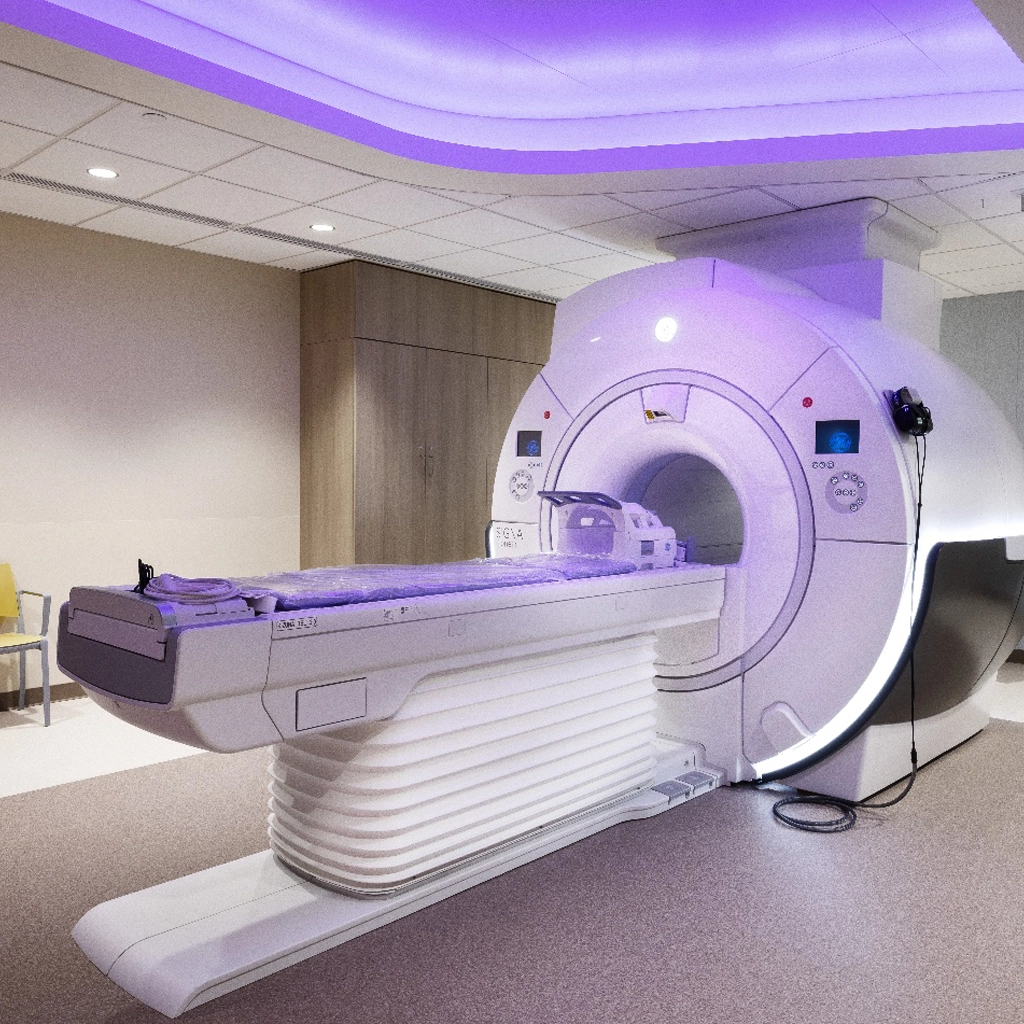
An MRI machine looks similar to a CT scanner. It features a table for patients that moves in and out of a large tube. Depending on the part of the body being studied, you may be placed feet or head first on the table. Some patients get nervous because the inside of the MRI scanner is tight on all sides, but the test is perfectly safe and the table can be easily moved if needed.

All imaging studies require a written MRI prescription or referral stating the exam being ordered and the medical necessity of the procedure. An MRI exam lasts 20 to 60 minutes depending on the body part being scanned and is usually divided into multiple short imaging segments.

During each segment, you’ll hear a series of loud noises, often described as intermittent high-pitched banging. You will be provided a pair of headphones to reduce the noise. During the scan, you’ll need to stay as still as possible. To make sure doctors get a clear picture, you may also be asked to hold your breath for a short period of time.

In some instances, a contrast agent (gadolinium DTPA) will be administered when requested by the referring physician.

For patients with claustrophobia, IV sedation is available at our East Brunswick office on request. We also offer high-field open MRIs (MRI machines with no closed tunnel), which patients may find less stressful. Arrangements can also be made for the patient to speak to our nurses and visit the office prior to the exam to see the equipment and learn about the scanning procedure.

What Happens After an MRI?

Patients are usually discharged shortly after completing the MRI procedure. Then the images are sent to a specialized doctor called a radiologist. The radiologist looks at the images and provides a detailed report to your physician. You may receive a phone call from your doctor to discuss your MRI results, or schedule a follow-up appointment.

For more information about Magnetic Resonance Imaging (MRI) at University Radiology Group, or to schedule an appointment with one of our specialists, please contact us.