Diagnostic X-ray Procedures at University Radiology Group

X-Rays vs. Other Scans | Common Types | Women’s Health | Preparation | What To Expect | Results

What Is a Diagnostic X-ray?

Diagnostic X-ray procedures are studies that use small amounts of radiation to visualize different parts of your body. During these tests, X-ray beams pass through the body and produce an image on a special medical imaging film or digital program. Some X-ray imaging tests use contrast agents like barium or iodine to enhance the images.

x-ray

Generally, the benefits of X-rays outweigh the risks associated with radiation. However, X-ray tests do carry some risk of pregnancy issues. Always tell your radiology care team if you are or may be pregnant before an X-ray.
Some patients can also have allergic reactions to IV contrast agents. Symptoms of an allergic reaction include lightheadedness, hives, itching, and nausea. In rare cases, severe reactions can cause difficulty breathing and/or swelling.

walk-in x-ray

X-Rays vs. Other Scans

X-ray studies are often used to help diagnose bodily injuries, blockages in the body, and certain tumors and illnesses. For example, X-rays can be used to identify broken bones, assess the severity of pneumonia in the lungs, and detect bowel obstructions. Additionally, dental X-rays are commonly used to track the position and health of a patient’s teeth.

When compared to other imaging studies, X-rays are faster than MRIs and use less radiation than CT scans. However, while X-rays are useful for highlighting musculoskeletal issues, they are not the preferred test for soft-tissue concerns like stroke or appendicitis.

Common Types of X-ray Tests

Bone & Joint Studies

  • Arthrogram: Visualizes cartilage, ligaments and any loose bodies in a joint to highlight tears, defects, and other issues. The study requires an injection of contrast or contrast and air into the joint space.
  • Bone Density Scan (DXA): Studies the bones (usually the spine and hips) to check for osteoporosis (bone loss).

Barium Studies (Fluoroscopy)

  • Barium Enema: Looks for structural issues, inflammation, and signs of colon cancer in the large intestine using barium and air introduced through the rectum. Patient preparation is required prior to the test.
  • Esophagram (Barium Swallow Test): Highlights issues in the esophagus, such as problems swallowing and hiatal hernias after the patient drinks a barium solution.
  • Small Bowel Series: Helps doctors find anatomical issues and inflammation in the small intestine and requires the patient to drink a barium solution.
  • Upper GI Series: Uses multiple studies of the esophagus, stomach and the beginning of the small intestine to diagnose issues in the GI tract. Patients need to drink a barium solution with gas granules before the test.
  • UGI and Small Bowel: Visualizes the esophagus, stomach and entire small intestine, and requires the patient to drink a barium solution with gas granules.

Bladder and Kidney X-Rays

  • Cystogram: Uses contrast material delivered to the bladder through a catheter to assess bladder health.
  • VCUG (Voiding Cystourethrogram): Checks for urinary issues with the help of contrast material. After filling the bladder with a contrast solution, pictures are taken as the patient urinates.

Myelogram

Myelograms study the spinal column and nerves. Under fluoroscopic guidance, the radiologist places a needle into the spine (“spinal tap”). They inject contrast using the same catheter before obtaining X-ray images. Myelogram scans help doctors identify nervous system problems such as pinched nerves, herniated disks, and spinal stenosis.

Women’s Health X-rays

Hysterosalpingogram

This specialized X-ray visualizes the uterus and fallopian tubes with the help of contrast injected through the cervical opening. Hysterosalpingograms are often used to diagnose fertility issues by assessing uterus shape, determining fallopian tube patency, and checking for fibroids and polyps.

Mammography (Digital Mammogram)

A mammography is an X-ray study used to detect lesions in breast tissue. They can show potential tumors even before a mass can be felt. This makes mammography particularly valuable as a screening tool in detecting early breast cancer.

Mammograms are a routine procedure for women beginning at the age of 40. Younger women may be prescribed mammograms if they have a family history of breast cancer or carry certain genetic markers.

Some patients report discomfort during a mammogram as the breasts are compressed. However, the compression is not dangerous and does not damage breast tissue. In fact, it improves the accuracy of your mammogram and reduces the amount of radiation needed for the test. Scheduling your mammogram within the first 14 days after your period can reduce discomfort.

Routine mammograms at University Radiology Group are interpreted by Mammography Specialists at the University Radiology Digital Screening Mammography Program.

Other Diagnostic X-ray Studies

X-rays can also be used to visualize injuries and illnesses within the bones, head, chest, spine, abdomen, or extremities. They can assist with diagnosis and help doctors assess improvement after surgery, physical therapy, or other treatments.

Preparing for an X-ray Study

All diagnostic X-ray exams require a written prescription or referral stating which exam is being ordered and why. If the X-ray will involve a contrast agent, you’ll be screened for asthma, allergies, diabetes, and any use of an inhaler before your appointment. If you are diabetic, you’ll also need to provide a complete list of medications.

If you’ve had prior X-rays in the same part of the body, we typically request a copy of those studies for comparison. This information will expedite the report of the diagnosis back to the referring physician.

Some procedures may have additional preparation instructions. If preparation is required, you can download our Adult or Pediatric Prep Sheet Instructions or get a copy at one of our offices.

What To Expect During a Diagnostic X-ray

Before bringing you to the X-ray machine, you’ll be asked to change into a medical gown. You’ll also need to remove all jewelry, and may be asked to remove bras with underwire, as metal can interfere with X-ray images.

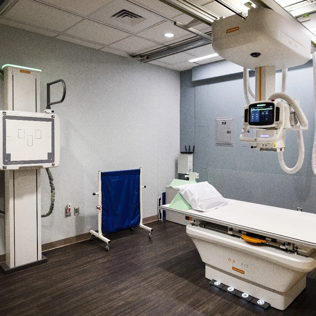
The X-ray technician will then position you for your X-ray study. Depending on the part of the body being scanned, you may be asked to sit, stand, or lie down on a medical table. In many cases, they’ll give you lead pads to protect your reproductive organs.

To keep pediatric patients still and in the correct position for their X-ray exam, technicians sometimes use “pediatric immobilization devices.” Common devices include special chairs with moving parts and straps, tubing that holds a child’s arms above their head, and special swaddles for infants.

The technician will go into a separate observation room before taking the X-ray images. You’ll need to stay as still as possible, and may be asked to hold your breath for scans of your torso. If multiple images are needed, the X-ray technician will come into the room between images to change your position. Most X-ray studies take 10-15 minutes.

Getting Your X-Ray Results

After your X-rays are complete, films are interpreted by a specialized doctor called a radiologist. The radiologist creates an in-depth report of the images, including the size and location of any issues. Then they send the report and/or films to your referring physician, who will follow up with you on the results.

Schedule your X-ray appointment at our comfortable, convenient outpatient clinic »

For more information about Diagnostic X-ray Procedures at University Radiology Group, or to schedule an appointment with one of our specialists, please contact us.