Women’s Imaging

Overview | Services | Breast Density | Subspecialty Expertise and Why it Matters

The most comprehensive women’s imaging services in New Jersey
All women’s imaging providers are not the same! There are differences in the equipment used and with the experience and training of the doctors who interpret women’s imaging tests.

When you have your screening or diagnostic tests performed at one of our imaging centers or at one of the medical centers we support, you benefit from the region’s leading experts on the issues that affect a woman’s health. This includes breast cancer screening and diagnostic imaging, as well as bone density screening for an issue that affects women to a four times greater degree than men – osteoporosis.

We also employ 26 radiologists who are fellowship (subspecialty) trained in women’s imaging. This added expertise gives our patients and the physicians who care for them an added measure of confidence in this rapidly advancing and constantly improving field.

Our women’s imaging services include:
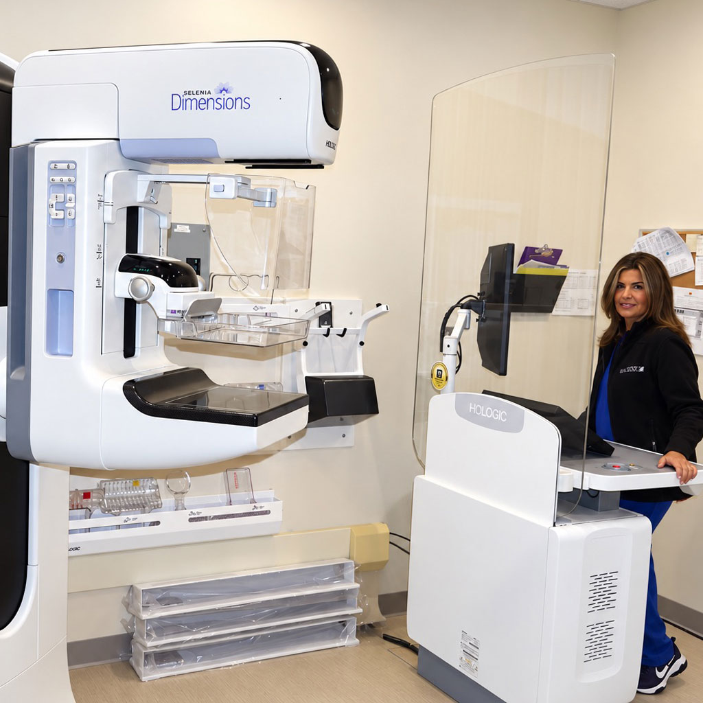
Mammo Tech
  • Advanced and 3D mammography: The most advanced digital breast tomosynthesis screening for better detection with fewer false positives, as well as extensive diagnostic capabilities.
  • Breast ultrasound: A diagnostic test that uses high-frequency sound waves to create detailed images of breast tissue, typically used to evaluate lumps or abnormalities found on a mammogram.
  • Breast MRI: An imaging test that uses a powerful magnet, radio waves and a computer to create detailed images of breast tissue. Can be used to detect and evaluate breast abnormalities.
  • Breast biopsy: We offer several different methods for performing breast biopsy, depending on the initial test that detected the abnormality. The biopsy is used to take a sample of breast tissue for further evaluation in a laboratory.
  • Ultrasound-guided breast cyst aspiration: An imaging-guided procedure that uses a thin needle to remove cyst fluid for diagnosis or to relieve symptoms.
  • Bone densitometry (DXA): A low-dose x-ray test to measure bone mineral density to assess bone strength and help diagnose conditions such as osteoporosis.

Breast Density

What is breast density?

Breasts are made up of a mixture of fibrous and glandular tissue and fatty tissue. Your breasts are considered dense if you have a lot of fibrous or glandular tissue but not much fat. Density may decrease with age, but there is little, if any, change in most women.

How do I know if I have dense breasts?

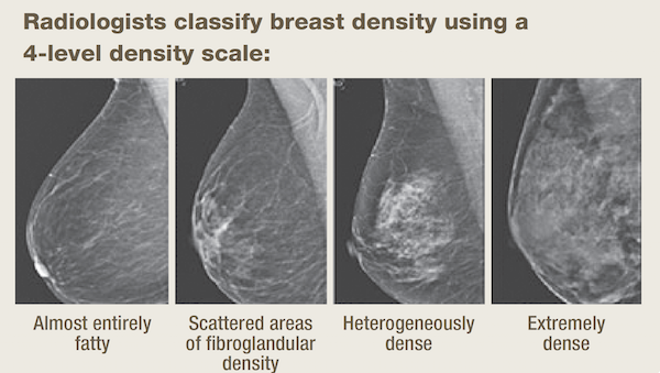
Breast density is determined by the radiologist who reads your mammogram. There are four categories of mammographic density. The radiologist assigns each mammogram to one of the categories. Your doctor should be able to tell you whether you have dense breasts based on where you fall on the density scale. (See scale)

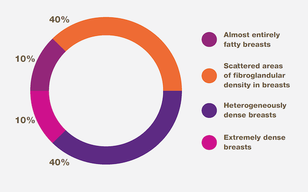
Breast Density in the U.S. (See chart)

  • 10% of women have almost entirely fatty breasts
  • 10% have extremely dense breasts
  • 80% are classified into one of the two middle categories

Why is breast density important?

Having dense breast tissue may increase your risk of getting breast cancer. Dense breasts also make it more difficult for doctors to spot cancer on mammograms. Dense tissue appears white on a mammogram. Lumps, both benign and cancerous, also appear white. So, mammograms can be less accurate in women with dense breasts.

Are there any tests that are better than a mammogram for dense breasts?

In breasts that are dense, cancer can be hard to see on a mammogram. Studies have shown that ultrasound and magnetic resonance imaging (MRI) can help find breast cancers that can’t be seen on a mammogram. However, both MRI and ultrasound, show more findings that are not cancer, which can result in added testing and unnecessary biopsies. Also, the cost of ultrasound and MRI may not be covered by insurance.

Subspecialty expertise and why it matters

The term “subspecialty” refers to the optional, additional fellowship training that a radiologist can undertake in a specific area of diagnostic imaging. This is especially important in women’s imaging because this field involves complex imaging techniques, nuanced interpretation and conditions where early and accurate diagnoses can significantly affect outcomes.

Subspecialty expertise can contribute to:

  • Improved cancer detection: Subspecialty expertise can improve the detection of subtle findings and reduce the chance of missed cancers.
  • More accurate interpretation: Abnormal findings on a mammogram can be challenging to distinguish between benign and malignant. Subspecialty-trained radiologists are trained to understand variations in breast anatomy, identify high-risk lesions and know the difference in appearance of different cancer types.
  • Fewer false positives: The added training of a subspecialized radiologist can help patients avoid unnecessary additional testing and procedures.
  • Keeping pace with advancing technology: Because women’s imaging is a rapidly evolving field, subspecialists stay abreast of advancements specific to breast health. This can help them leverage new equipment and techniques that contribute to earlier and more accurate diagnoses.

University Radiology employs 26 doctors with subspecialty (fellowship) training in women’s imaging. By offering greater expertise in this area, women can be assured that the most highly-trained doctors are reading and interpreting their screening and diagnostic imaging tests.

For more information about Women’s Imaging at University Radiology Group, or to schedule an appointment with one of our specialists, please contact us.