Breast Biopsy at University Radiology Group

Overview | Why is a breast biopsy performed? | Types | Preparation | What to Expect | Test Results | Frequently Asked Questions

Breast Biopsy Overview

If there is a suspicious finding seen on a breast imaging test, your doctor may order a breast biopsy to determine whether or not the finding is cancerous. During a breast biopsy, a sample of cells or tissue from the area of concern is taken and sent to a specialized doctor called a pathologist.

Breast biopsies are outpatient procedures performed by a trained breast radiologist using a needle with image guidance, such as mammography, ultrasound, or MRI.
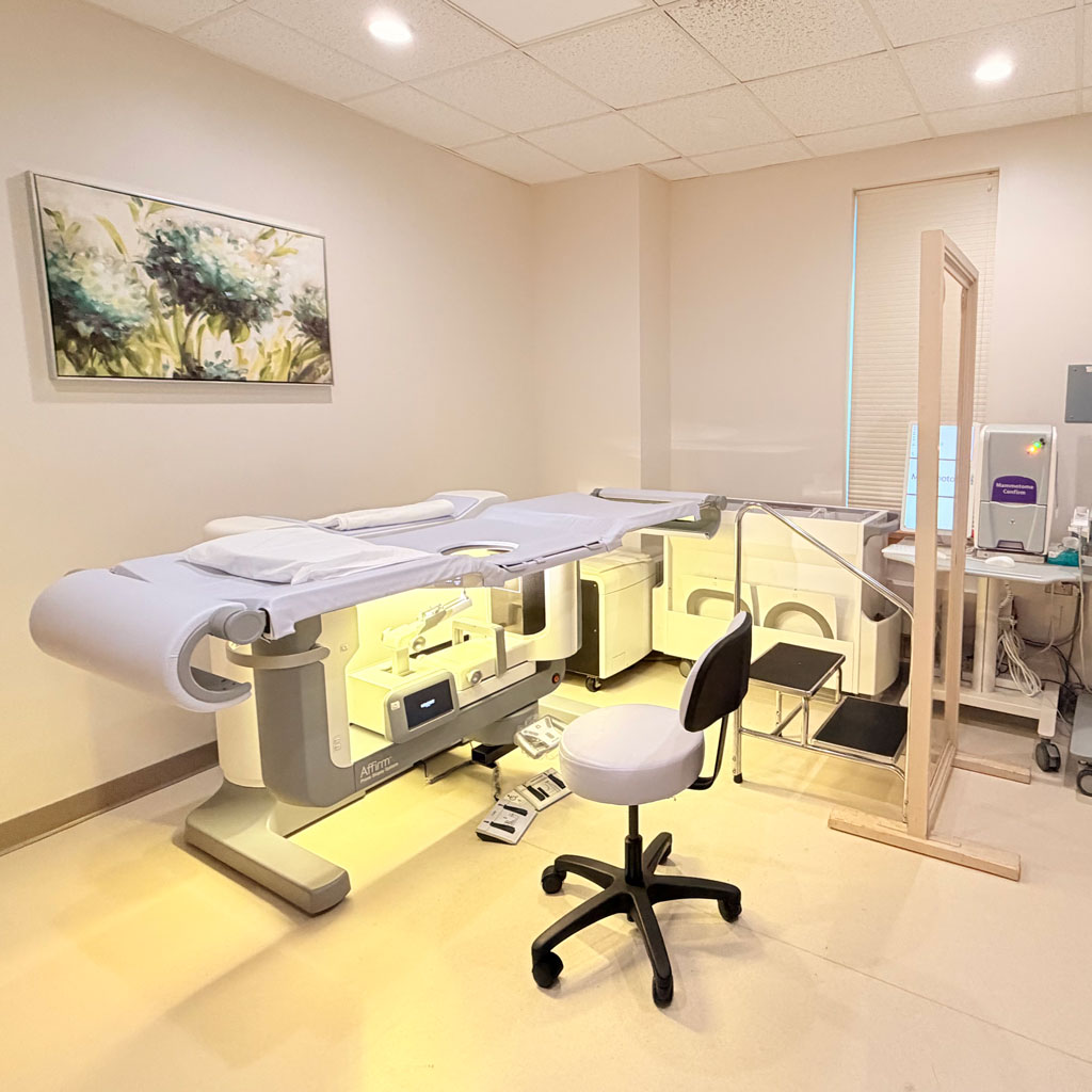

Stereotactic Breast Biopsy Room

Why is a breast biopsy performed?

A breast biopsy is performed to examine a breast abnormality more closely, such as a mass, calcification, or area seen on a mammogram, ultrasound, or MRI. A small sample of breast tissue is removed to determine whether the abnormality is benign (noncancerous) or cancerous. A biopsy is the only way to definitively diagnose breast cancer.

Types of Breast Biopsies

Sterotactic

A sterotactic biopsy uses a vacuum assisted needle and mammographic images to target the abnormality.

MRI

An MRI uses a vacuum assisted needle and MRI images to target the abnormality.

Ultrasound

An ultrasound uses a vacuum assisted needle or core needle and ultrasound images to target the abnormality.

How do I know which breast biopsy is right for me?

The right type of breast biopsy depends on how the area of concern is best seen and how it appears on your breast imaging. Ultrasound-guided biopsy is commonly used when the abnormality can be clearly seen on ultrasound; stereotactic biopsy uses mammography to target areas such as calcifications; and MRI-guided biopsy is recommended when an abnormality is best seen on MRI. Your radiologist will review your imaging and recommend the biopsy approach that provides the most accurate way to evaluate the area.

What to Expect During a Breast Biopsy

  • You will undress from the waist up and put on a gown.
  • You will be awake and will receive a local anesthetic to minimize discomfort.
  • A breast radiologist will perform your biopsy.
  • No stitches are needed.
  • We will send the samples to a lab for analysis.
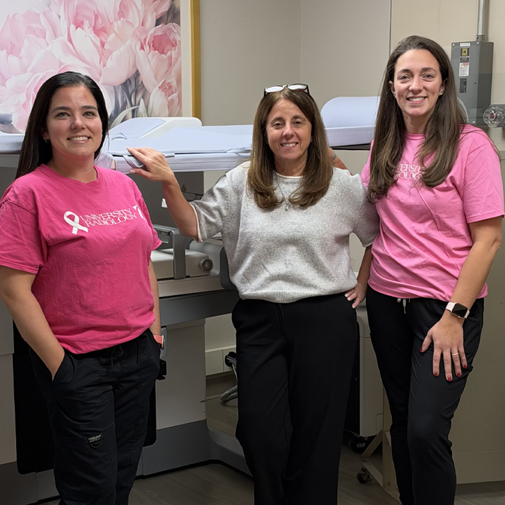
Dr. Lina Paster, Krista Olesinski and Ashley Collins

Featured are: Dr. Lina Paster (center), Clinical Section Chief of Mammography and our Mammography clinical coordinators Krista Olesinski and Ashley Collins

What to expect for each type of breast biopsy:

Mammogram (stereotactic) core needle biopsy

  • You will lie face down on a movable exam table, and your breast will be positioned and compressed, similar to a mammogram. You may need to remain in this position for approximately 30–60 minutes.
  • Mammogram images will be taken to locate the area of concern and guide the biopsy.
  • You will receive a local anesthetic to numb the area of the breast to be biopsied.
  • The breast radiologist will make a small incision, approximately 1/4 inch long. No stitches are typically required.
  • Using additional mammogram images to guide precise placement, the radiologist will insert the biopsy needle and remove multiple tissue samples.
  • A small marker clip will be placed at the biopsy site to identify the location for future imaging.
  • The tissue samples will be sent to a laboratory for analysis.

MRI guided vacuum-assisted biopsy

  • A nurse or technologist will insert an intravenous (IV) line into a vein in your hand or arm to administer contrast dye, which helps identify the area of concern.
  • You will lie face down on the MRI table, and your breast will be positioned and held in place using a specialized grid.
  • MRI images will be taken to locate the area of concern and guide the biopsy.
  • You will receive a local anesthetic to numb the area of the breast to be biopsied.
  • The breast radiologist will make a small incision, approximately 1/4 inch long. No stitches are typically required.
  • Using additional MRI images to guide precise placement, the radiologist will insert the biopsy needle and use a vacuum-assisted device to remove multiple tissue samples.
  • A small marker clip will be placed at the biopsy site to identify the location for future imaging.
  • The tissue samples will be sent to a laboratory for analysis.
  • You may need to remain in this position for approximately 30–60 minutes during the procedure.

Ultrasound-guided core needle biopsy

  • You will lie face up or slightly turned to the side on an exam table.
  • Ultrasound images will be taken to locate the area of concern and guide the biopsy.
  • You will receive a local anesthetic to numb the area of the breast to be biopsied.
  • The breast radiologist will make a small incision, approximately 1/4 inch long. No stitches are typically required.
  • Using ultrasound images to guide precise placement, the radiologist will insert the biopsy needle and remove multiple tissue samples.
  • A small marker clip will be placed at the biopsy site to identify the location for future imaging.
  • The tissue samples will be sent to a laboratory for analysis.

Frequently Asked Questions

You may feel some breast discomfort and/or pinching and burning from the numbing medication and pressure during the procedure. Some soreness, bruising, or swelling afterward is common. If you experience significant or worsening pain during or after your biopsy, please inform us immediately.

A small marker clip will be placed at the biopsy site after the tissue samples are collected. The clip helps identify the exact location of the biopsy for future mammograms or other breast imaging.

No, in fact most breast biopsies do not result in a cancer diagnosis. But since the biopsy is the only reliable way to determine what an abnormal finding actually represents, it is important to have one to make sure.

Your results can range from a normal (or benign) breast tissue to non-cancerous conditions. If cancer is found, the pathology report may provide additional information about the type, grade and hormone-receptor or HER2 status.

Most pathology results are available within a few business days, although some take longer if additional testing or specialist review is necessary.

The breast radiologists at University Radiology have received additional training in various aspects of breast imaging. They are among the region’s leading experts, having performed more than 3,000 breast biopsies last year at 10 different locations.

The following 10 locations perform breast biopsies.

For more information about breast biopsy at University Radiology Group, or to schedule an appointment with one of our specialists, please contact us.