
3D Digital Mammograms at University Radiology Group

2D vs. 3D Mammography | Artificial Intelligence + Expert Breast Imaging | Experience in Breast Imaging | Who Needs a Digital Mammogram? | Preparation | What to Expect | Results | FAQ

3D Mammography Overview

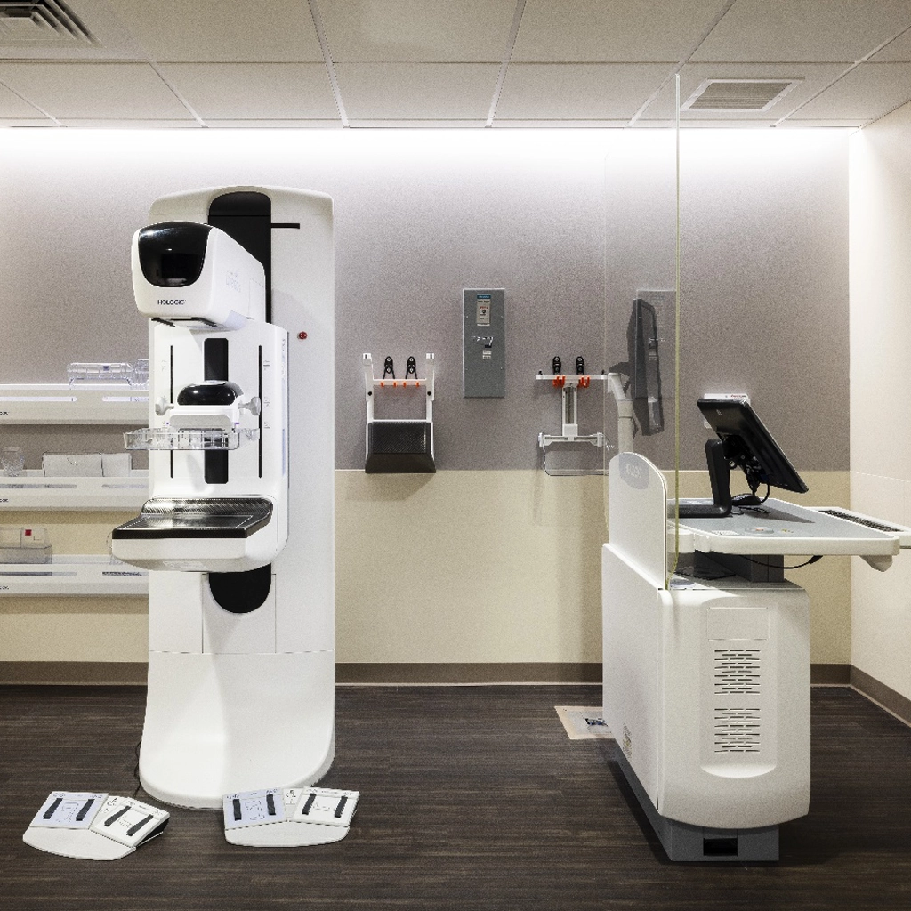
Digital mammography, also known as full-field digital mammography or digital breast tomosynthesis (DBT), is a specialized X-ray study of the breasts. It combines 3D and traditional 2D imaging to build a finely detailed 3D reconstruction of breast tissue. Radiology physicians can then scroll through all the images much like a book – one page at a time – to gain a better understanding of your breast health.

mammogram

3D mammography is used for both breast cancer screenings and as a diagnostic tool. Because it recreates three-dimensional images of the breast tissue, digital mammograms can often detect cancerous tumors before they can be felt. This makes digital mammography particularly valuable as a screening tool in the early detection of breast cancer.

The test is FDA approved and recommended for patients in all risk categories.

2D vs. 3D Mammography

Sometimes breast tissue can overlap. When viewed only with conventional 2D mammography, overlapping areas may look like abnormalities. This can lead to a false positive result that requires patients to get additional testing, increases their healthcare costs, and creates unnecessary anxiety.

Examining multiple images with 2D and 3D techniques allows radiologists to view tissue more clearly, making it easier to make an accurate assessment. Additionally, the 3D images make it possible to identify cancers at earlier stages when they are most treatable.

Who Needs a Digital Mammogram?

The American Cancer Society recommends that women with average risk of breast cancer begin getting annual mammograms at the age of 40. Women with an elevated risk of breast cancer should get screening mammography starting at 30 years old. These risks include:

  • Family history of breast cancer
  • Mutation of the BRCA1 or BRCA2 gene
  • An immediate family member with a BRCA1 or BRCA2 gene mutation
  • Previous radiation therapy of the chest
  • Other genetic conditions such as Li-Fraumeni syndrome

In some high-risk cases, a breast MRI is recommended in addition to a mammogram.

Preparing for a 3D Mammogram

There is no special preparation for a digital mammography exam. However, patients should not use any powders, creams or deodorant under the arms or around the breast area before the exam.

As part of the complete exam, current films are always compared with prior studies. Patients should bring any prior mammogram studies with them for their appointment. Please note that it is very important that all mammograms are returned to us so that the patient has a complete history file.

What to Expect During a Mammography Exam

Digital mammograms use similar equipment to traditional 2D mammograms. When you get to your mammography appointment, you’ll first change into a medical gown. A radiology technician will then bring you to an X-ray room with a mammography machine. The technician will position your breasts on a support plate and lower a second plate down to compress the breast tissue. Then they’ll go into an adjoining room, from which they can operate the X-ray machine.

During 3D image acquisition, the X-ray arm will move in an arc over the breast, taking multiple X-ray images over the course of a few seconds. The technologist will view the images to ensure they are of the highest quality. The exam usually takes less than 15 minutes.

All of our offices are equipped with the latest digital mammography equipment. Schedule your digital mammogram today by filling out an online form or calling us at (732) 390-0033.

Mammogram Exam Results

Routine screening digital mammograms are interpreted by Digital Mammography Specialists at University Radiology. If additional views are needed to make the diagnosis, our office will contact you to return for specialized spot or magnification views.

If no additional images are needed, the radiologist will send a detailed report to your referring physician. Your doctor will call or schedule an appointment to discuss the results with you.

Frequently Asked Questions

Some women find the compression required for a mammogram uncomfortable. Women with dense breast tissue or small breasts often report more discomfort, as firmer compression may be needed to provide accurate breast imaging. It’s important to remember that the compression is completely safe and will not damage your breast tissue. Any discomfort from the procedure is temporary and can be managed with oral pain medication. Scheduling your mammogram in the first 14 days after your period may result in a more comfortable experience.

Digital mammograms, 3D mammography, and digital breast tomosynthesis all refer to the same imaging test.

With the more advanced, lower dose C-ViewTM software, the radiation exposure required for the 2D together with the 3D exam is now about the same as with a traditional 2D mammogram alone.

A screening mammogram is a routine evaluation of the breast tissue. A diagnostic mammogram focuses on a concerning area of the breast or is used to evaluate a symptom or complaint. 2D and 3D mammography can be used together in both cases.

Many insurance companies cover 3D mammograms, but it’s best to check with your insurance provider to ensure coverage and understand your health care policy.

For more information about 3D Digital Mammograms at University Radiology Group, or to schedule an appointment with one of our specialists, please contact us.